How
should I prepare for my MRI procedure?
How should I prepare for my MRI procedure?
The magnetic field used for MRA will pull on any
iron-containing object in the body, such as a heart pacemaker, intrauterine
device, vascular access port, metal plate, pins, screws or staples. You
will be given a questionnaire to answer regarding these issues. The radiologist
or technologist should know about any such item and also whether you have
ever had a bullet in your body, whether you ever worked with metals, or
if you have had a joint replacement. If there is any question, an x-ray
can be taken to detect metal objects.
The radiologist should also know if you have fillings
in your teeth, which could distort images of the facial region or brain.
Braces make it harder to properly adjust the MRI unit. You will be asked
to remove hairpins, jewelry, eyeglasses, hearing aids and any dental work
that can be taken out. Some wigs contain metal and must be removed. Red
dyes used in tattoos and permanent eyeliner may contain metallic iron, but
this is rarely a problem. You should report any drug allergies to the radiologist
or technologist and should mention if there's any possibility that you might
be pregnant.
You can eat normally before the exam (unless told
differently), but a young child should not eat or drink for about four hours
if they will receive a sedative. The rules vary at different MRI facilities,
so be sure to check with your medical center about eating and drinking before
the exam. Medications may be taken as usual. Some patients will feel uncomfortably
confined (claustrophobic) when enclosed in an MRI unit. If necessary, you
will be given a sedative to help put you at ease, though probably fewer
than one in every 20 patients will need this. You will wear a lightweight
medical gown for the exam.
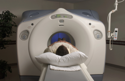
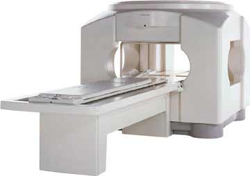
What does the equipment look like?
The traditional MRI unit is a large tube surrounded
by a circular magnet, in which the patient lies without moving for several
seconds at a time. The patient is placed on a wheeled bed that is moved
into the magnet. In recent years, patient-friendly units have been designed,
and examination in such units is becoming increasingly available. These
machines are both shorter and wider than a conventional MRI unit and do
not fully enclose the patient. Some of the newer C-shaped units are even
open on all sides and are thus very attractive to patients who tend to be
claustrophobic. A drawback is that image quality is not as consistently
good.
Example of the MRI equipment that may be used
is shown to the left.
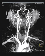
How does the procedure work?
Exposing the patient to radio waves in a strong
magnetic field generates data that are used by a computer to create images
of tissue slices that may be viewed in any plane or from any direction.
The magnetic field lines up atomic particles in the tissues called protons,
which are then spun by a beam of radiofrequency waves and produce signals
that are picked up by a receiver in the imager. It is these signals that
are processed by the computer to produce images. The resulting images are
very sharp and detailed and are thus able to demonstrate tiny changes from
the normal pattern that are caused by disease or injury. Special settings
are used to image various structures, such as arteries in the case of MRA.
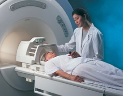
How is the procedure performed?
The patient is placed on a special table and positioned
inside the opening of the MRI unit. A typical exam consists of two to six
imaging sequences, each taking two to 15 minutes. Each sequence provides
a specific image orientation and a specified degree of image clarity or
contrast. Depending on the type of exam being done, the total time needed
can range from 10 to 60 minutes, not counting the time needed to change
clothing, have an IV put in and answer questions. When contrast material
is needed, a substance called gadolinium is given by IV injection during
one of the imaging sequences. It highlights blood vessels, making them stand
out from surrounding tissues.
The radiologist and technologist leave the
examining room during the actual imaging process, but the patient can communicate
with them at any time using an intercom. Some centers permit a friend to
stay nearby, or a parent if a child is being examined. When the exam is
completed you will be asked to wait to make sure that more images are not
needed.
What will I experience during a MRI procedure?
The technologist will make you as comfortable as possible, but at times
the magnet may be within a few inches of your face. For those who become
very uncomfortable when enclosed in a small space, a mild sedative is nearly
always effective. You may notice a warm feeling in the area being studied.
This is normal, but do not hesitate to report it if it bothers you. If you
receive a contrast material injection, there may be some local discomfort
at the IV site. The loud tapping or knocking noises that are heard during
certain parts of the exam disturb some patients; earplugs may help.
Who interprets the MRI results and how do I get them?
A radiologist experienced in MRI will analyze
the results and send a report to your physician, along with an interpretation
of the findings. Your physician in turn will discuss the MRA findings with
you. Some centers now send diagnostic reports and images over the Internet,
speeding up the process.
What are the benefits vs. risks?
Benefits
- Detailed images of blood vessels and blood flow
are obtained without having to insert a catheter directly into the area
of interest, so that there is no risk of damaging an artery.
- The procedure itself and the time needed to
recover are shorter than after a traditional catheter angiogram.
- MRA is less costly than catheter angiography.
- There is no exposure to x-rays during an MRI
study.
- Even without using contrast material, MRA can
provide high-quality images of many blood vessels, making it very useful
for patients prone to allergic reactions.
- As with catheter-based angiography or CT angiography,
it frequently is possible to defer surgery after getting the results of
an MRA study. If surgery remains necessary, it can be performed more accurately.
Risks
- There are no definite side effects from any type
of MRI study, including MR angiography. Claustrophobia may be a problem,
however. When it is severe and not relieved by giving a sedative, an alternative
imaging method may have to be tried. If a metal implant is present but
goes undetected, it may be affected by the strong magnetic field to which
the patient is exposed. In addition, if the implant is close to the examination
site it may be hard to get high-quality images.
- MRI is generally avoided during the first three
months of pregnancy. Ultrasound is preferred at this time, unless the
woman might have a very serious condition that is best detected with MRA.
The effects of MRI on the fetus, if any, remain to be determined. The
general rule for MRI and other diagnostic studies in pregnancy is that
they should be avoided unless there is substantial risk from missing the
correct diagnosis because the procedure is not done. Women who are breast-feeding
should inform the radiologist and ask how to proceed. They may pump breast
milk before the exam for use until the gadolinium contrast material has
cleared from the body.