Welcome
to Global Diagnostic Imaging OnLine Educational
Resource. As the name connotes, this area of our site will provide additional
information and resources related to topics in CT, MRI Ultrasound as well
as other medical resources.
This section of our website is a mix of information
you can use in your daily practice, along with news about cutting-edge diagnostic
and therapeutic techniques.
We welcome suggestions for future content
or other ways we can improve understanding of medical conditions.
Imaging
Education
To
View the latest Cardiovascular Update Click on the Image of the Newsletter
Mayo
Publication Update
MRI
Safety Concerns
Maybe you're concerned about the long-term impact of having all your atoms
mixed about, but once you're out of the magnetic field, your body and its
chemistry return to normal. There are no known biological hazards to humans
from being exposed to magnetic fields of the strength used in medical imaging
today.
How
MRI Works
What
Is Ultrasound
Ultrasound
(US) imaging, also called ultrasound scanning or sonography, is a method of
obtaining images from inside the human body through the use of high frequency
sound waves. The reflected sound wave echoes are recorded and displayed as
a real-time visual image. Ionizing radiation (x-rays) are not involved in
ultrasound imaging.
Ultrasound
is a useful examination tool for many of the body's internal organs, including
the heart, liver, gallbladder, spleen, pancreas, kidneys, and bladder. Because
ultrasound images are captured in real-time, they can show movement of internal
tissues and organs, and enable physicians to see blood flow and heart valve
functions.
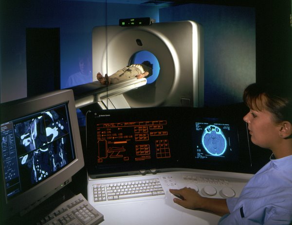
How
MRI Works -
MRI
scanners vary in size and shape, and some newer models have a greater degree
of openness around the sides. Still, the basic design is the same, and the
patient is pushed into a tube that's only about 24 inches (60 centimeters)
in diameter. But what's in there?
What
is Ultrasound
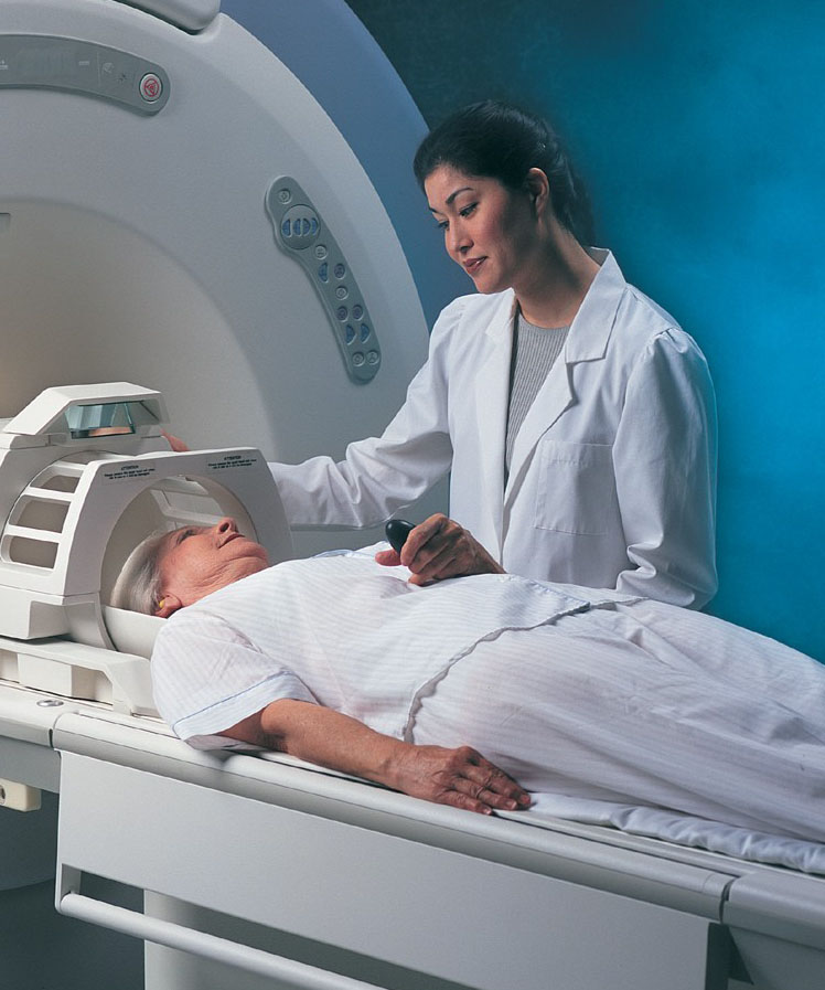
MRI
Safety Concerns -
The
biggest and most important component of an MRI system is the magnet. There
is a horizontal tube -- the same one the patient enters -- running through
the magnet from front to back. This tube is known as the bore. But this isn't
just any magnet -- we're dealing with an incredibly strong system here, one
capable of producing a large, stable magnetic field.
To
View the latest Clinical Update Click on the Image of the Newsletter
Medical
Links
Media
Presentations
Please
note: if you experience any problems
with the playback of any video components within our web site you may need
the latest version of a media player. We have included a quick links for you
to download them. (see below)..
ABI
Exam Importance & Demonstration
5
minute presentation about the importance and a demonstration of how to perform
an ABI test.
PLAYHeart
Sounds
Heart
Sound Tutorial that tests your auscultation skills. Extra sounds, diastolic
and systolic murmurs in recordings of actual patients
PLAYMedia
Players:
To
download media software Select the player you wish to install. Click on the
link. You will be directed to their web site. Follow instructions for downloading
and installing media software.
Resources
Ankle-Brachial
Index - (ABI) result is used to predict
the severity of peripheral arterial disease (PAD).
Intima-Medial
Thickness -(IMT), also called intimal
medial thickness, is a measurement of the thickness of artery walls to track
the progression of atherosclerotic disease.
Framingham
Risk - Framingham coronary prediction
algorithm provides estimates of total CHD risk.
What
Is Diastolic Dysfunction - Patient
Information - explains the process of loss of elasticity that causes stiffening
of the heart and how it can be treated.