

How does the procedure work?
Ultrasound imaging is based on the same physical principles involved in the sonar used by bats, ships at sea, and anglers with fish detectors. As the sound passes through the body, the transducer produces and records echoes that can identify how far away an object or organ is, how large it is, its shape, and its consistency (fluid, solid or mixed).
The ultrasound transducer functions as both a generator of sound (like a speaker) and a detector (like a microphone). When the transducer is pressed against the skin, it directs inaudible, high-frequency sound waves into the body.
How is the procedure performed?
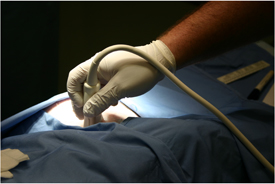
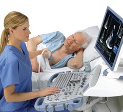
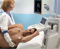
The sonographer or technologist usually spositions the patient on an examination table. A clear gel applied to the patient's body in the area to be examined helps the transducer make secure contact with the skin. The sound waves produced by the transducer cannot penetrate air, so the gel helps eliminate air pockets between the transducer and the skin. The technologist presses the transducer firmly against the skin and sweeps it back and forth to image the area of interest.
When the examination is complete, the patient can dress. The technologist can review the ultrasound images in real time as they are acquired, and the patient can be released immediately. Imaging results are then forwarded to the physician for further analysis and review.

What is General Ultrasound Imaging?
Ultrasound (US) imaging, also called ultrasound scanning or sonography, is a method of obtaining images from inside the human body through the use of high frequency sound waves. The reflected sound wave echoes are recorded and displayed as a real-time visual image. Ionizing radiation (x-rays) are not involved in ultrasound imaging. Obstetric ultrasound refers to using sound waves to visualize an embryo or fetus in the uterus of a pregnant woman thus determine the condition of either mother or child.

What are the limitations of General Ultrasound Imaging?
Ultrasound has difficulty penetrating bone and other dense materials. For this reason, ultrasound can only depict the outer surface of bony structures and not what lies within. For visualizing bone, your physician may recommend other imaging modalities such as magnetic resonance imaging (MRI).
What are the benefits vs. risks?
Benefits

What are some common uses of the procedure?
Millions of expectant parents have seen the first "picture" of their unborn child with ultrasound examinations of the fetus. Ultrasound imaging is used extensively for evaluating the eyes, pelvic and abdominal organs, heart, and blood vessels, and can help a physician determine the source of pain, swelling, or infection in many parts of the body.
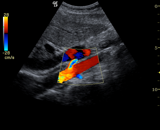
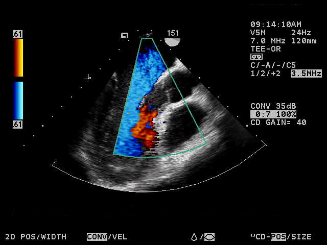
Doppler ultrasound is a special technique used to examine blood flow. Doppler images can help the physician to see and evaluate:
What will I experience during the procedure?
Most ultrasound examinations are painless, fast, and easy. You will lie on your back or side on an examining table. The technologist will spread some gel on your skin and then press the transducer firmly against your body, moving it until the desired images are captured. There may be varying degrees of discomfort from pressure as the technologist guides the transducer over the area being examined. The examination usually takes less than 60 minutes.
How should I prepare for the procedure?
You should wear comfortable, loose-fitting clothing for your ultrasound exam. Other preparation depends on the type of examination you will have.
Risks
Who interprets the results and how do I get them?
A
cardiologist, experienced in ultrasound and other procedures, will analyze
the images and submit a signed report with his/her interpretation to the
patient’s personal physician. The patient receives a copy of the ultrasound
results from the referring physician who ordered the test. In some cases
the technologist may discuss preliminary results with you at the conclusion
of your examination with your referring physician. SonoNet's digital imaging
technology also allows for distribution of diagnostic reports and referral
images over the Internet. Most SonoNet facilities are equipped for this.
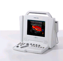
What does the equipment look like?
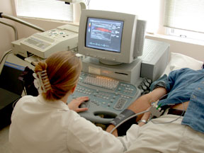
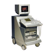
Ultrasound scanners consist of a console containing a computer and electronics, a video display screen and a transducer that is used to scan the body. The transducer is a small, hand-held device about the size of a bar of soap, attached to the scanner by a cord. The technologist spreads a lubricating gel on the patient in the area being examined, and then presses the transducer firmly against the skin to obtain images.
The ultrasound image is immediately visible on a nearby screen that looks much like a computer or television monitor. The physician or technologist watches this screen during an examination and captures representative images for storage. Often, the patient is able to see it as well.