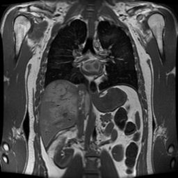
What is MRI Angiography?
Magnetic resonance imaging (MRI) is a method
of producing extremely detailed pictures of body tissues and organs without
the need for x-rays. The electromagnetic energy that is released when exposing
a patient to radiofrequency waves in a strong magnetic field is measured
and analyzed by a computer, which forms two- or three-dimensional images
that may be viewed on a TV monitor. MR angiography (MRA) is an MRI study
of the blood vessels. It utilizes MRI technology to detect, diagnose and
aid the treatment of heart disorders, stroke, and blood vessel diseases.
MRA provides detailed images of blood vessels without using any contrast
material, although a special form of contrast material is often given to
make the MRI images even clearer. The procedure is painless, and the magnetic
field is not known to cause tissue damage of any kind.
What
is MRI Angiography?
What are some common uses of a MRI procedure?
MRI is commonly used to:
- Many patients with arterial disease now have
it treated in the radiology department rather than undergoing surgery
in an operating room. MRA is a very useful way of finding problems with
blood vessels and determining how to best to treat those problems.
- The carotid arteries in the neck that conduct
blood to the brain are a common site of atherosclerosis, which may severely
narrow or block off an artery, reducing blood flow to the brain and even
causing a stroke. If an ultrasound study shows that such disease is present,
many surgeons will perform the necessary operation after confirmation
with MRA, dispensing with the need for catheter angiography.
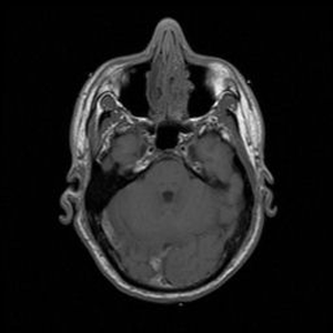
- MRA has found wide use in checking patients
for diseased intracranial (in the head) arteries, so that only those with
positive findings will need to undergo a more invasive catheter study.
- MRA is also used to detect disease in the aorta
and in blood vessels supplying the kidneys, lungs and legs.
- Patients with a family history of arterial aneurysm,
a ballooning out of a segment of the vessel wall, can be screened with
MRA to see if they have a similar disorder that has not produced symptoms.
If an aneurysm is found, it may be eliminated surgically, possibly avoiding
serious or fatal bleeding.
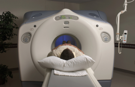
How should I prepare for a MRI procedure -
The
magnetic field used for MRA will pull on any iron-containing object in the
body, such as a heart pacemaker, intrauterine device, vascular access port,
metal plate, pins, screws or staples. You will be given a questionnaire to
answer regarding these issues. The radiologist or technologist should know
about any such item and also whether you have ever had a bullet in your body,
whether you ever worked with metals, or if you have had a joint replacement.
If there is any question, an x-ray can be taken to detect metal objects.